
Post-COVID-19 pneumothorax, secondary to ruptured pneumatocele or bulla?
DOI:
https://doi.org/10.32818/reccmi.a8n1a2Keywords:
anaesthesia, bullae, COVID-19Abstract
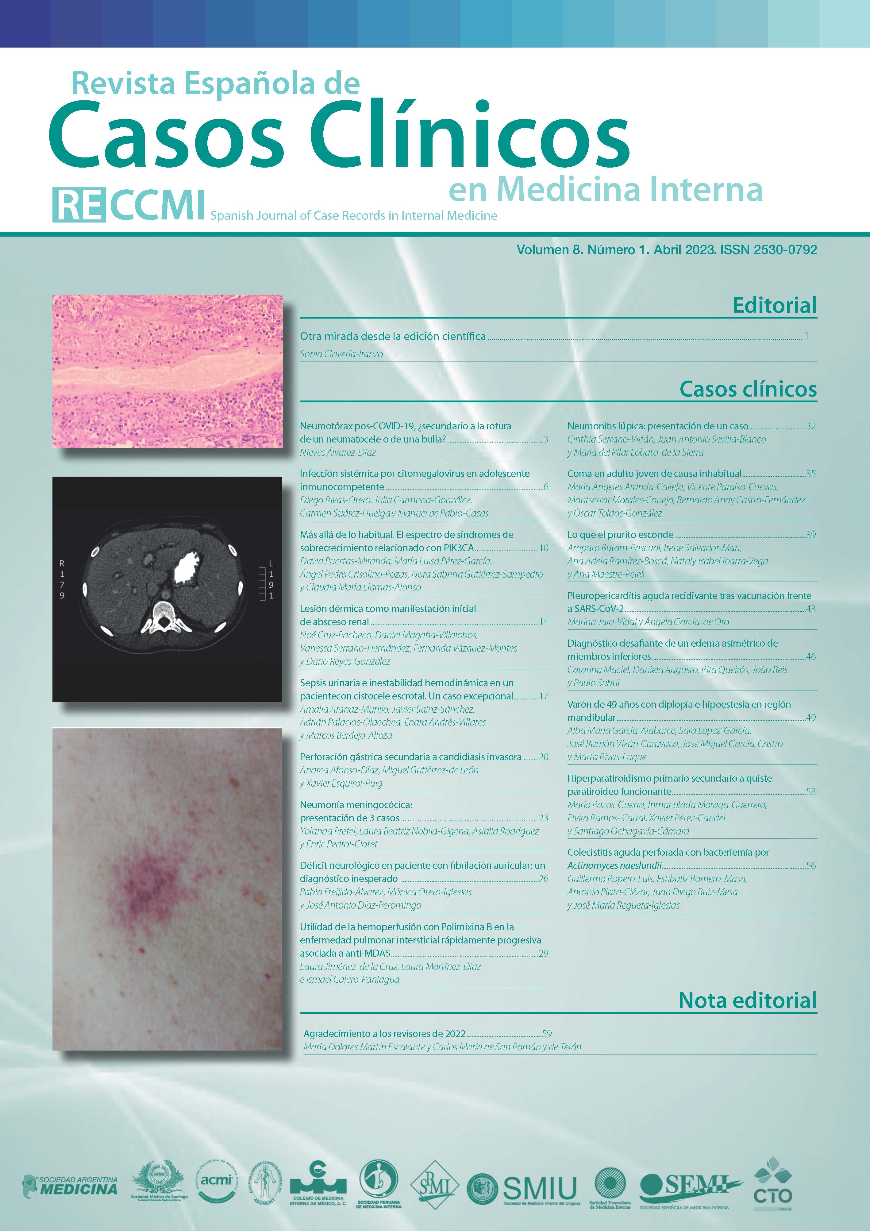
Pneumatoceles in patients with COVID-19 have been described in the literature however, little is known about its incidence and patophisiology. Clinically and with available imaging techniques is hard to discern between a pneumatocele and a bullae. Both have different phisiopatology and treatment, the final diagnosis is always anatomopathological. We report the perioperative management of a 41 year old man scheduled for right thoracotomy, aerostasis , neumatocele resection and pulmonary absess by Staphilococcus Aureus Meticillin sensitive after COVID-19 pneumonia. The perioperative course was without incident. The anatomophatologic analysis reveled a subpleural bullae and intense fibrosis.
Downloads
Metrics
References
McCann C, Shoeib M,Rashid MI, Kostoulas N. Pneumatocele formation following COVID-19 pneumonia. Is there a role for surgical intervention. Asian Cardiovasc Thorac Ann. 2021; 30(4): 2184923211059866. doi: https://doi.org/10.1177/02184923211059866 (último acceso ene. 2023). DOI: https://doi.org/10.1177/02184923211059866
Jolobe OMP. Air leaks, pneumatoceles, and air spaces in Covid-19 pneumonia. Am J Emerg Med. 2021; 46: 785. doi: https://doi.org/10.1016/j.ajem.2020.08.098 (último acceso ene. 2023). DOI: https://doi.org/10.1016/j.ajem.2020.08.098
Chang SH, Chen D, Paone D, Geraci TC, Scheinerman J, Bizekis C et al. Thoracic surgery outcomes for patients with Coronavirus Disease 2019. J Thorac Cardiovasc Surg. 2021; 162(6): 1654-1664. doi: https://doi.org/10.1016/j.jtcvs.2021.01.069 (último acceso ene. 2023). DOI: https://doi.org/10.1016/j.jtcvs.2021.01.069
Hamad AM, El-Saka HA. Post COVID-19 large pneumatocele: clinical and pathological perspectives. Interact Cardiovasc Thorac Surg. 2021; 33(2): 322-324. doi: https://doi.org/10.1093/icvts/ivab072 (último acceso ene. 2023). DOI: https://doi.org/10.1093/icvts/ivab072
Wu J, Feng LC, Xian XY, Qiang J, Zhang J, Mao QX et al. Novel coronavirus pneumonia (COVID-19) CT distribution and sign features. Zhonghua Jie He He Hu Xi Za Zhi. 2020; 43(4): 321-326. Chinese. doi: https://doi.org/10.3760/cma.j.cn112147-20200217-00106 (último acceso ene. 2023).
Schiller M, Wunsch A, Fisahn J, Gschwendtner A, Huebner U, Kick W. Pneumothorax with Bullous Lesions as a Late Complication of Covid-19 Pneumonia: A Report on Two Clinical Cases. J Emerg Med. 2021; 61(5): 581-586. doi: https://doi.org/10.1016/j.jemermed.2021.04.030 (último acceso ene. 2023). DOI: https://doi.org/10.1016/j.jemermed.2021.04.030
Sun R, Liu H, Wang X. Mediastinal Emphysema, Giant Bulla, and Pneumothorax Developed during the Course of COVID-19 Pneumonia. Korean J Radiol. 2020; 21(5): 541-544. doi: https://doi.org/10.3348/kjr.2020.0180 (último acceso ene. 2023). DOI: https://doi.org/10.3348/kjr.2020.0180
Downloads
Published
How to Cite
Issue
Section
License
Copyright (c) 2023 Nieves Álvarez-Díaz

This work is licensed under a Creative Commons Attribution-NonCommercial-NoDerivatives 4.0 International License.
Permite compartir, copiar y redistribuir el material en cualquier medio o formato, bajo los siguientes términos:
Reconocimiento: debe otorgar el crédito correspondiente, proporcionar un enlace a la licencia e indicar si se realizaron cambios. Puede hacerlo de cualquier manera razonable, pero no de ninguna manera que sugiera que el licenciante lo respalda a usted o su uso.
No comercial: no puede utilizar el material con fines comerciales.
No Derivados: si remezcla, transforma o construye sobre el material, no puede distribuir el material modificado.
Sin restricciones adicionales: no puede aplicar términos legales o medidas tecnológicas que restrinjan legalmente a otros de hacer cualquier cosa que permita la licencia.


